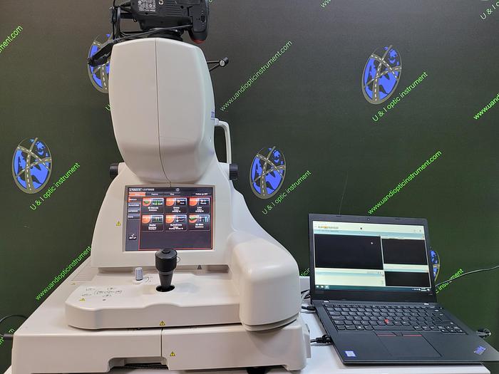
2011 Topcon 3D OCT-2000 FAplus

2011 Topcon 3D OCT-2000 FAplus
€13,950 (EUR)
Location:Netherlands
Description
Description
The TOPCON 3D OCT-2OOO Series is an optimal choice for all eye care professionals
The 3D OCT-2000 Series of Spectral Domain OCTs with High Resolution Fundus Cameras was designed to meet the needs of a comprehensive fundus imaging device for all eye care professionals from the single doctor practice to a large
>> OCT, Color, Red-free*1, FA, FAF images acquirable
OCT Technology with the Integrated Fundus Camera
12 x 9 mm wide scan & 5 line cross scan!
Rich analysis functions plus high resolution images
50,000 A-scans/sec – Greater details in shorter time
The enhanced 50,000 A-scans/sec allows for faster tomography acquisition and available to produce clear crosssectional retinal images. Now there are even more imaging variations with the new 12×9mm wide scan enabling the user to capture a wider area of the retina from optic disc to macula with a single shot. Additionally the 5 Line Cross Scan can be a perfect solution for detailed screening and quick follow-up. Moreover, Topcon’s “ Enhanced Choroidal Mode ” visualizes further internal structures, allowing much superior visualization of the interface between choroid and sclera. Data analysis is now selectable from 2 formats – Fine or Basic, and can be performed fast or in detail according to your purposes. Experience sophisticated examination with the new evolved Topcon 3D OCT-2000 Series.
Stunning retinal images with integrated high resolution retinal camera
Combining OCT and a color fundus camera in one unit, the Topcon 3D OCT-2000 line-up is perfected now with FA and FAF photography functions. Furthermore digital Red-free images can be displayed easily at the touch of a button. Owing to flexibly changeable ISO sensitivity, reduced flash level with crystal-clear fundus observation is available resulting in reduced patient fatigue and miosis. If the OCT image is only required, simply select “Color Photography OFF”.
» Anterior segment analysis
Corneal thickness map, corneal thickness distribution
diagram, curvature radius distribution diagram, curvature
radius and peripheral corneal thickness analysis, manual
angle measurement are all available.
- In order to capture anterior segment photography, it is necessary to use the headrest attachment.
FA – FAF
* Import function
FA, FAF, ICG, Red-free images can be imported easily
into Fastmap software. Simultaneous observation of OCT
and imported images become available.
Specifications
| Manufacturer | Topcon |
| Model | 3D OCT-2000 FAplus |
| Year | 2011 |
| Condition | Refurbished |
| Stock Number | 1703427 |


